

When pain travels into your buttocks or legs, or when numbness and tingling make everyday activities difficult, even walking a short distance or getting a good night's sleep can feel like a challenge. Over time, these symptoms may begin to affect your independence, confidence, and quality of life.
For some people, the underlying cause is lumbar foraminal stenosis, a condition in which the openings through which spinal nerves exit the lower back become narrowed. When a nerve is compressed, it can cause pain, weakness, or altered sensation that interferes with daily routines and makes it difficult to stay active.
If conservative treatments have not provided enough relief, you may be wondering whether surgery could help. Understanding the available lumbar foraminal stenosis surgery options is an important step toward making an informed decision about your spine health. The right procedure depends on where the nerve is compressed, how severe your symptoms are, and whether your spine remains stable.
Eexploring your spine care options and understanding what factors may influence a surgical recommendation. With the right information from Dr Navarro and an individualized assessment, you can approach your next steps with greater clarity and confidence.
What Is Lumbar Foraminal Stenosis?
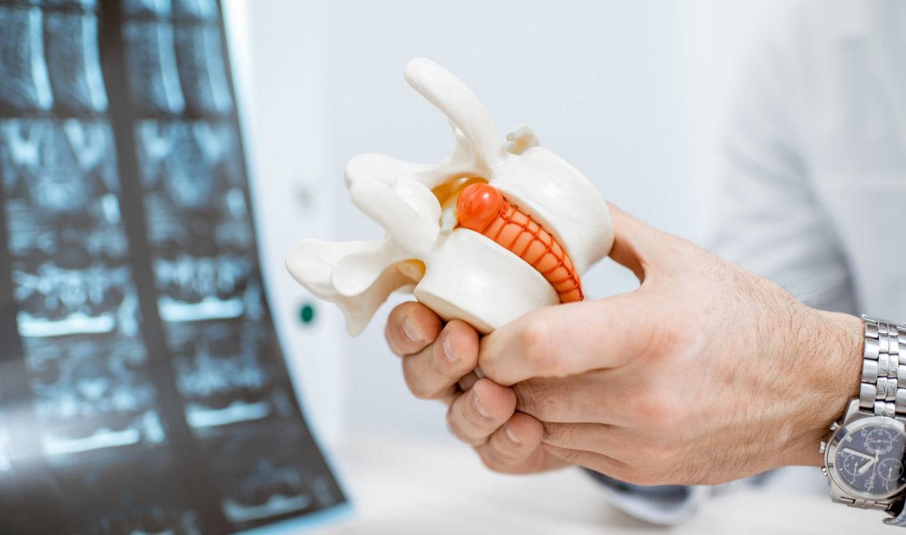
The lumbar spine consists of five vertebrae in the lower back. Between these vertebrae are intervertebral discs that provide cushioning and help the spine move. Small openings called neural foramina are located on either side of the spine, allowing spinal nerves to exit and travel to other parts of the body.
Lumbar foraminal stenosis develops when one or more of these openings become narrower, leaving less room for the nerves. As the available space decreases, a nerve root may become irritated or compressed.
What Causes Foraminal Narrowing?
Several changes can contribute to lumbar foraminal stenosis. Age related degeneration is a common factor, as spinal discs gradually lose height and flexibility. When a disc becomes thinner or bulges outward, the space around the existing nerve may decrease.
Bone spurs, also called osteophytes, can develop as the spine changes over time. Thickened ligaments, arthritic changes in the facet joints, and a herniated disc may also contribute to narrowing.
In some cases, spondylolisthesis, a condition in which one vertebra slips relative to another, changes the alignment of the spine and reduces the available space for a nerve.
The underlying cause matters because different anatomical problems may require different surgical approaches.
What Symptoms Can Lumbar Foraminal Stenosis Cause?
Symptoms depend on the affected nerve root and the degree of compression. Some people experience lower back pain, while others mainly notice pain that radiates into the buttock, thigh, calf, or foot.
Numbness, tingling, burning sensations, and muscle weakness can also occur. These symptoms may worsen during standing, walking, or certain movements, although the pattern varies from person to person.
The severity of symptoms does not always match the degree of narrowing seen on an MRI. A surgeon must consider imaging findings alongside your medical history, symptoms, and physical examination to determine whether the narrowed foramen is responsible for your problems.
When Is Surgery Recommended for Lumbar Foraminal Stenosis?
Surgery is not the first treatment for every person with foraminal stenosis. Many patients begin with nonsurgical approaches, particularly when symptoms are manageable and there is no significant or progressive neurological deficit.
Treatment may include physical therapy, activity modification, appropriate medication, and selected spinal injections. These approaches aim to reduce symptoms and improve function, although they do not necessarily reverse the structural narrowing.
When Conservative Treatment Is Not Enough
A surgical consultation may be appropriate when persistent nerve pain limits walking, sleeping, working, or other daily activities despite an adequate trial of nonsurgical treatment.
Surgery may also be considered when examination and imaging findings indicate significant nerve compression that corresponds with the patient's symptoms.
When Neurological Symptoms Require Prompt Assessment
Progressive weakness deserves timely medical evaluation. New difficulty lifting the foot, worsening leg weakness, or increasing problems with balance may indicate significant nerve dysfunction.
Sudden loss of bladder or bowel control, numbness around the groin or saddle area, or rapidly progressing leg weakness requires emergency medical assessment. These symptoms may indicate a serious condition, such as cauda equina syndrome, and should not be managed by waiting for a routine consultation.
Lumbar Foraminal Stenosis Surgery Options
The primary goal of surgery is to create enough space for the affected nerve to exit the spine without harmful compression. The best approach depends on the location and cause of the narrowing, the extent of bone or disc changes, and whether removing tissue could compromise spinal stability.
1. Foraminotomy, Enlarging the Nerve Opening
A foraminotomy is a procedure designed to widen the neural foramen and relieve pressure on an exiting spinal nerve. The surgeon removes selected portions of bone or other obstructing tissue that restrict the opening.
Depending on the anatomy, this may involve removing part of the facet joint, a bone spur, or tissue contributing to the narrowing. The objective is to decompress the nerve while preserving as much of the normal spinal structure as possible.
Foraminotomy may be appropriate when the compression is primarily located in the neural foramen and the spine is sufficiently stable.
The procedure can be performed using different surgical techniques, including conventional open surgery or minimally invasive approaches. The most suitable method depends on the patient's anatomy, the extent of compression, and the surgeon's assessment.
2. Lumbar Decompression Surgery
Lumbar decompression is a broader term for procedures that remove or reshape structures pressing on spinal nerves. A decompression procedure may include a foraminotomy, removal of part of the lamina, or removal of a portion of a disc when that tissue contributes to nerve compression.
A laminotomy involves removing a small portion of the lamina, the bony covering at the back of the spinal canal, to improve access to compressed neural structures.
A laminectomy removes a larger portion of the lamina and is commonly used when stenosis affects the central spinal canal. It may be combined with foraminal decompression when narrowing occurs in multiple areas.
However, a laminectomy alone may not adequately address isolated foraminal stenosis. The surgical plan must target the specific location where the nerve is compressed.
Decompression can be effective for appropriately selected patients, particularly when symptoms are caused by mechanical nerve pressure and the spine does not require stabilization.
3. Minimally Invasive Foraminal Decompression
Minimally invasive spine surgery uses specialized instruments and smaller incisions to reach the affected structures while limiting disruption to surrounding tissues.
For selected cases of lumbar foraminal stenosis, the surgeon may use a tubular retractor, magnification, or an endoscope to remove bone and other tissue compressing the nerve.
Potential advantages include less muscle disruption, reduced blood loss, and a shorter hospital stay or recovery period for some patients. However, these benefits depend on the specific procedure, the patient's health, and the complexity of the spinal condition.
Minimally invasive surgery is not automatically the best option for every patient. Severe narrowing, extensive bony changes, complex anatomy, or spinal instability may require a different approach.
The priority is to achieve adequate nerve decompression safely, not simply to make the incision as small as possible.
4. Discectomy for Foraminal Stenosis Caused by a Herniated Disc
A herniated disc occurs when disc material extends beyond its normal boundaries and may press against a nearby nerve root. When a herniation contributes to foraminal narrowing, removing the offending disc material may relieve compression.
A discectomy involves removing the portion of the disc responsible for pressing on the nerve. In selected cases, the surgeon may perform a microdiscectomy using magnification and a limited surgical exposure.
This approach is most relevant when a herniated disc is an important cause of the patient's symptoms. If bone spurs, facet joint enlargement, or disc height loss are the main reasons for foraminal stenosis, a discectomy alone may not create sufficient space.
The surgical plan must therefore address the actual source of nerve compression rather than relying on a single procedure for every patient.
5. Spinal Fusion When Instability Is Present
Spinal fusion is different from decompression. Instead of simply removing tissue that presses on a nerve, fusion joins two or more vertebrae to limit movement between them.
A surgeon may consider fusion when foraminal stenosis occurs alongside significant spinal instability, certain cases of spondylolisthesis, deformity, or other structural problems. Fusion may also be considered when adequate decompression would require removing enough of a stabilizing facet joint to create a meaningful risk of instability.
By stabilizing the affected spinal segment, fusion may help maintain alignment and support the surgical result. Depending on the case, an interbody fusion technique may also help restore disc height, potentially increasing the space available for the exiting nerve.
However, spinal fusion is not necessary for every patient with lumbar foraminal stenosis. It is a more extensive intervention that can involve longer recovery, risks associated with bone healing, and changes in the mechanical demands placed on neighboring spinal segments.
The decision should be based on a clear clinical reason to stabilize the spine, not simply the presence of stenosis on an MRI.
How the Location of Nerve Compression Influences Surgery
Lumbar foraminal stenosis can affect different nerve roots and may occur at one or multiple spinal levels. Identifying the precise location of compression is essential for selecting the appropriate procedure.
Foraminal Versus Extraforaminal Compression
Foraminal compression occurs within the opening where the nerve exits the spine. In this situation, a targeted foraminotomy may provide the necessary space.
Extraforaminal compression occurs farther outside the foramen, sometimes because of a disc herniation, bone spur, or other structural change. This may require a surgical approach that reaches the nerve beyond the usual exit opening.
Central canal stenosis is different because it affects the main passageway containing the spinal nerves. When central and foraminal narrowing occur together, the surgeon may need to address both areas.
Single Level Versus Multilevel Stenosis
Some patients have narrowing at a single spinal level, while others have several affected levels. A single level problem may allow for a more focused operation.
Multilevel stenosis requires careful planning because the surgeon must determine which narrowed areas are actually responsible for the symptoms. Treating every abnormality seen on imaging is not always necessary or beneficial.
The aim is to address the clinically significant compression while preserving healthy structures whenever possible.
What Factors Determine the Best Surgical Option?
Choosing among lumbar foraminal stenosis surgery options requires more than reviewing an MRI. A surgeon must understand how the anatomy, symptoms, and overall health of the patient interact.
Symptom Severity and Neurological Function
Persistent radiating leg pain, numbness, and functional limitations can support consideration of surgery when they correspond with the imaging findings.
Progressive muscle weakness may increase the urgency of assessment. The surgeon will evaluate whether the nerve compression is likely to improve with decompression and whether any neurological changes may be permanent.
Spinal Stability and Alignment
The stability of the spine is one of the most important factors in deciding whether decompression alone is appropriate.
If the affected segment is stable, a targeted procedure may relieve nerve pressure without fusion. If there is significant instability or a risk of creating instability during decompression, the surgeon may consider stabilization.
MRI Findings and Physical Examination
MRI can show disc changes, bone spurs, narrowing of the neural foramina, and possible nerve root compression. However, imaging findings must be interpreted in context.
A surgeon may also assess strength, sensation, reflexes, walking ability, and other clinical findings. Additional tests may be appropriate if symptoms and MRI results do not clearly identify the source of the problem.
Age, General Health, and Recovery Goals
Other considerations include overall health, bone quality, smoking status, other medical conditions, previous spinal operations, and the patient's goals.
For example, a patient who hopes to return to walking longer distances may have different functional priorities from someone whose main concern is persistent pain while sitting or sleeping.
Discussing these priorities with your surgeon can help establish realistic expectations and a treatment plan that reflects your needs.
What Are the Risks and Recovery Expectations After Surgery?
All spinal procedures carry potential risks, and the details vary according to the operation performed.
Possible complications include infection, bleeding, blood clots, dural tear with cerebrospinal fluid leakage, nerve injury, and persistent or recurrent symptoms. Decompression may not eliminate all pain, particularly if another condition contributes to the symptoms.
Fusion procedures have additional considerations, including failure of the bones to unite, hardware complications, and stress on adjacent spinal levels.
Recovery After Lumbar Decompression
After decompression, patients are generally encouraged to begin moving as directed by their surgical team. Some return home the same day, while others require a short hospital stay, depending on the procedure and their medical circumstances.
Leg pain caused by nerve compression may improve sooner than numbness or weakness. Nerves can take time to recover, and longstanding compression may leave some symptoms even after adequate decompression.
Recovery After Spinal Fusion
Recovery after fusion is often longer because the bones must heal together. Activity restrictions, follow up appointments, and rehabilitation recommendations vary according to the procedure and the patient's progress.
Your surgeon will explain when to resume driving, work, exercise, and other activities. Following postoperative instructions and attending follow up appointments are important parts of recovery.
Frequently Asked Questions About Lumbar Foraminal Stenosis Surgery
What is the most effective surgery for lumbar foraminal stenosis?
There is no single best procedure for every patient. A foraminotomy may be appropriate for isolated foraminal narrowing, while broader decompression may be needed when stenosis affects other parts of the spinal canal. Fusion may be considered when significant instability is present. The most effective option is the one that addresses the cause of compression while accounting for spinal stability and individual health needs.
Can lumbar foraminal stenosis be treated without surgery?
Yes. Depending on symptom severity, treatment may include physical therapy, activity modification, medication, and selected injections. These approaches can help manage symptoms, although they do not necessarily reverse the anatomical narrowing. Surgery may be considered when symptoms remain disabling despite appropriate nonsurgical care or when neurological deficits require more urgent intervention.
Is minimally invasive surgery better than open surgery for foraminal stenosis?
Minimally invasive techniques may reduce muscle disruption and help some patients recover more quickly. However, they are not suitable for every anatomical problem. The choice depends on the location and severity of compression, spinal stability, previous surgery, and the surgeon's ability to decompress the nerve adequately using the selected approach.
Does lumbar foraminal stenosis surgery require spinal fusion?
No. Many patients can undergo decompression without fusion if the spine is stable and the operation can preserve sufficient supporting structures. Fusion may be appropriate when instability, certain forms of spondylolisthesis, or the extent of necessary bone removal makes stabilization important.
Will surgery permanently relieve nerve pain?
Surgery can reduce symptoms caused by mechanical nerve compression, but results vary. Some patients experience substantial improvement, while others continue to have pain, numbness, or weakness. The outcome depends on factors such as the cause and duration of compression, the degree of nerve damage, other medical conditions, and whether the correct source of symptoms has been identified.
How do I know whether I need surgery for lumbar foraminal stenosis?
A specialist evaluation can help determine whether the narrowing seen on your MRI explains your symptoms. Your surgeon will consider your medical history, physical examination, imaging, response to nonsurgical treatments, and neurological function before discussing surgery. Seek emergency care if you develop sudden bladder or bowel dysfunction, saddle numbness, or rapidly progressing weakness.
Discuss Your Spine Care Options With Experts
Persistent lower back pain and radiating leg symptoms can make it difficult to enjoy everyday activities or feel confident about your future. When lumbar foraminal stenosis is responsible, understanding your options can help replace uncertainty with a clearer plan.
Lumbar foraminal stenosis surgery options range from targeted foraminotomy and minimally invasive decompression to discectomy and spinal fusion when stabilization is necessary. The right choice depends on where the nerve is compressed, how significantly your symptoms affect your life, and whether your spine is stable.
If you are struggling with ongoing symptoms or want to understand what your MRI findings mean, contact Dr Navarro to discuss your spine care options and ask whether a specialist evaluation is appropriate for your situation. Bring your imaging studies, radiology reports, and information about previous treatments so your concerns can be assessed in context.
Take the next step toward understanding your condition and making an informed decision about your spine health. With an individualized evaluation and a clear discussion of potential benefits and risks, you can explore the treatment approach that best fits your needs and recovery goals.

