
.svg)

Neck pain rarely begins as something alarming. It often starts quietly, a stiffness in the morning, a slight discomfort after a long day. But over time, that subtle pain can evolve into something more concerning, radiating sensations, weakness, or a loss of control that feels deeply unsettling.
For many patients, the uncertainty is the hardest part. You feel something is wrong, but you cannot see it. You cannot measure it. That is why the question what does a neck stenosis looks like becomes so important. It represents the moment when confusion turns into clarity.
Imaging provides that clarity. It transforms symptoms into something visible, something doctors can analyze and explain. For specialists like Dr. Navarro, these images are not just diagnostic tools, they are precise maps that guide every decision, from conservative care to advanced surgical solutions.
Understanding how cervical stenosis appears on imaging is not just about medical knowledge. It is about regaining control, making informed decisions, and taking the first step toward relief.
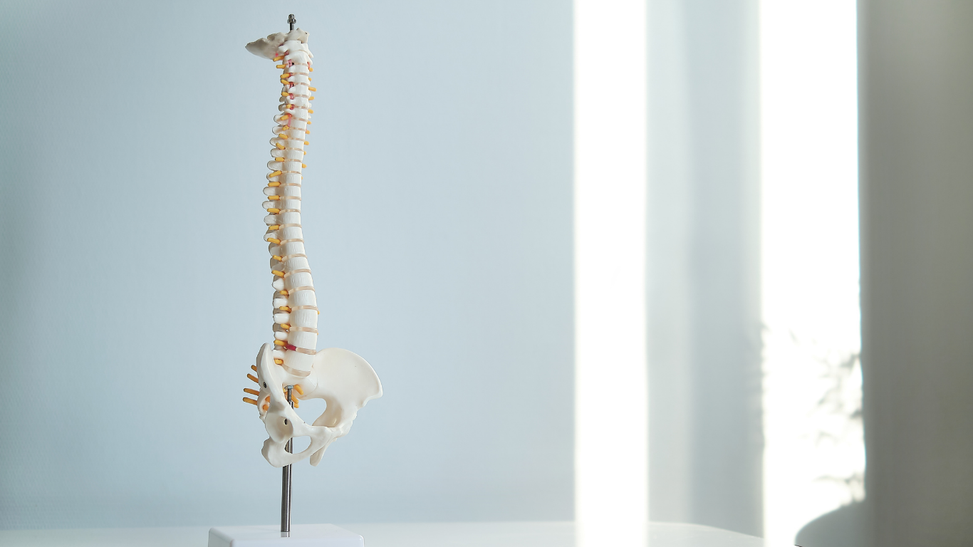
Understanding Cervical Stenosis
What Is Cervical Stenosis
Cervical stenosis is the narrowing of the spinal canal in the neck. This canal protects the spinal cord, which carries signals between your brain and body. When the space becomes reduced, the spinal cord or nearby nerves can become compressed.
This narrowing typically develops gradually. Age related degeneration is the most common cause, but disc herniations, thickened ligaments, and bone spurs can all contribute. Over time, these changes reduce the space available for critical neural structures.
Why Imaging Is Essential
Symptoms alone cannot confirm cervical stenosis. Many spine conditions present similarly, which makes imaging essential for an accurate diagnosis.
Imaging allows physicians to see inside the spine, identify the exact structures involved, and determine how severe the narrowing is. Without it, treatment decisions would rely on guesswork rather than precision.
What Does a Neck Stenosis Look Like on MRI
The Role of MRI in Diagnosis
Magnetic Resonance Imaging is the most important tool for evaluating cervical stenosis. It provides highly detailed images of soft tissues, including the spinal cord, intervertebral discs, and ligaments.
MRI is especially valuable because it shows not only the narrowing but also the effect on the spinal cord itself.
Key MRI Findings
On MRI, cervical stenosis appears as a visible reduction in the space surrounding the spinal cord. In a healthy spine, the cord is surrounded by a clear margin of space. In stenosis, that space becomes limited or disappears.
Doctors often identify disc bulges or herniations as one of the main causes. These appear as extensions of disc material pressing into the canal. Thickened ligaments can also be seen encroaching on the available space.
One of the most important findings is spinal cord compression. The cord may appear flattened or indented. In more advanced cases, there may be signal changes within the spinal cord, indicating irritation or damage.
What Severe Stenosis Looks Like
In severe cases, the spinal canal appears extremely tight. The spinal cord may be visibly compressed with little to no surrounding space. This is often associated with cervical myelopathy, a condition that can affect coordination, balance, and motor function.
MRI can also show whether multiple levels of the spine are involved, which plays a major role in determining the best treatment approach.
What Does Neck Stenosis Look Like on CT Scans
When CT Scans Are Used
Computed Tomography scans are primarily used to evaluate bone structures. While MRI focuses on soft tissues, CT provides a detailed view of the vertebrae and joints.
CT scans are often used when MRI is not possible or when additional detail about bony changes is required.
Key CT Findings
On a CT scan, cervical stenosis appears as a narrowing of the bony spinal canal. One of the most common findings is the presence of osteophytes, or bone spurs, which project into the canal.
Degenerative changes in the facet joints are also commonly seen. These changes can reduce the space available for the spinal cord and contribute to overall stenosis.
Comparing MRI and CT
MRI and CT scans complement each other. MRI shows how the spinal cord and soft tissues are affected, while CT reveals the structural and bony causes of the narrowing.
Together, they provide a complete understanding of the condition.
How Doctors Interpret Imaging Findings
Assessing the Degree of Narrowing
Doctors evaluate how much the spinal canal has narrowed. Mild stenosis may show slight crowding, while severe stenosis shows significant compression of the spinal cord.
This assessment is critical in determining the urgency and type of treatment required.
Identifying the Cause
Imaging helps identify the underlying cause of stenosis. It may be due to a disc herniation, bone spurs, ligament thickening, or a combination of factors.
Knowing the cause allows for more targeted and effective treatment.
Evaluating Spinal Cord Health
The condition of the spinal cord is one of the most important aspects of imaging interpretation. Evidence of compression or internal changes within the cord can indicate a more serious condition.
These findings often influence whether conservative treatment is sufficient or if surgical intervention is needed.
Common Imaging Terms You Might See in Your Report
Disc Bulge and Herniation
These terms describe disc material extending beyond its normal boundaries. They are common contributors to spinal canal narrowing.
Osteophytes
Osteophytes are bone spurs that develop due to degeneration. They can encroach on the spinal canal and reduce available space.
Canal Diameter Reduction
This refers to the measurable narrowing of the spinal canal. Reports may describe the degree of reduction to indicate severity.
Cord Compression
Cord compression means the spinal cord is being physically pressed. This finding is often associated with neurological symptoms.
Why Imaging Findings Matter for Treatment
Guiding Non Surgical Care
In less severe cases, imaging helps guide conservative treatments such as physical therapy, medications, or injections. The goal is to relieve symptoms without surgery.
Understanding the exact cause of stenosis allows for more precise and effective care.
Planning Surgical Intervention
When stenosis is severe or symptoms are worsening, imaging becomes essential for surgical planning. It helps determine which levels of the spine are affected and how best to relieve pressure on the spinal cord.
For Dr. Navarro, detailed imaging ensures that every intervention is carefully planned and tailored to the patient.
When Should You Get Imaging for Neck Pain
Warning Signs That Require Evaluation
Persistent neck pain, numbness, weakness, or coordination problems should not be ignored. These symptoms may indicate cervical stenosis or another serious condition.
Imaging provides the clarity needed to move forward with confidence.
The Importance of Early Diagnosis
Early diagnosis can prevent long term complications. Identifying cervical stenosis before significant damage occurs improves the chances of successful treatment.
Imaging is the key step in that process.
Frequently Asked Questions
What does a neck stenosis look like on an MRI scan
On MRI, neck stenosis appears as a narrowing of the spinal canal with reduced space around the spinal cord. In severe cases, the cord may appear compressed or flattened.
Can a CT scan detect cervical stenosis
Yes, CT scans can detect cervical stenosis, especially when caused by bone related changes such as osteophytes. However, they are less effective than MRI for evaluating the spinal cord.
Is imaging always necessary for neck pain
Not always. Mild, short term neck pain may not require imaging. However, persistent pain or neurological symptoms typically warrant further evaluation.
What is the difference between mild and severe stenosis on imaging
Mild stenosis shows limited narrowing with minimal impact on the spinal cord. Severe stenosis shows significant narrowing, often with clear spinal cord compression.
Take Control of Your Spine Health Today
Understanding what does a neck stenosis look like gives you more than information, it gives you direction. It allows you to move from uncertainty to clarity, from symptoms to solutions.
If you are experiencing ongoing neck pain or neurological symptoms, this is the moment to act. With the expertise of Dr. Navarro and advanced imaging techniques, you can receive a precise diagnosis and a treatment plan designed specifically for you.
Do not wait for the condition to progress. Take the next step, seek expert care, and begin your path toward lasting relief and improved quality of life.
.webp)